
Description
BioReady 150 nm Citrate-Stabilized Gold Nanoshells
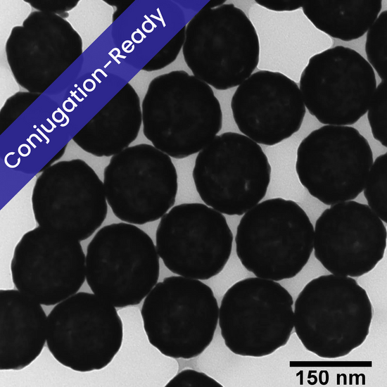
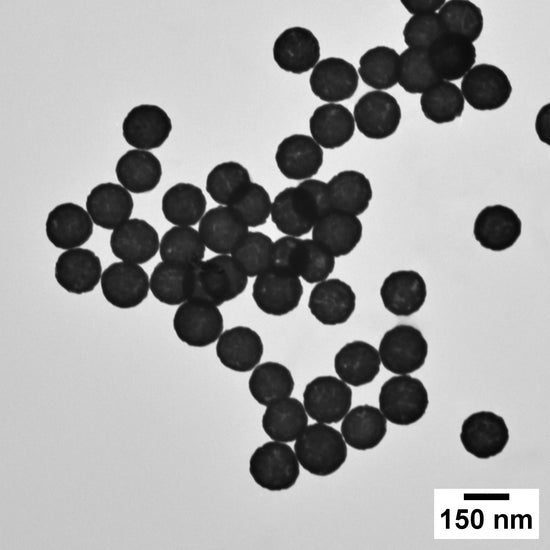
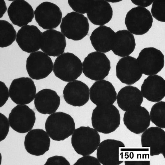
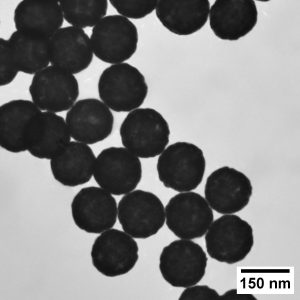
BioReady 150 nm Citrate-Stabilized Gold Nanoshells are core-shell plasmonic particles designed for high-sensitivity diagnostics, near-infrared optical detection, SERS, and photothermal research.
Each particle contains an amorphous silica dielectric core surrounded by a thin metallic gold shell. The core-shell ratio shifts the primary plasmon resonance into the near-infrared region, with an extinction maximum at approximately 770–810 nm.
Compared with conventional 40 nm solid gold nanospheres, these nanoshells provide a much larger optical extinction cross-section per particle. On lateral flow membranes, they produce distinct slate-blue to black lines, improving visual contrast for low-abundance analytes.
The particles are stabilized by a weakly bound sodium citrate layer in an aqueous 0.2 mM sodium citrate solution. The labile citrate coating supports passive adsorption of antibodies, proteins, and targeting peptides without requiring covalent coupling reagents.
Key features
- Near-infrared resonance: Engineered core-shell geometry provides an LSPR peak at approximately 770–810 nm.
- High optical extinction: The stated extinction cross-section is up to 30 to 100 times greater per particle than that of conventional 40 nm solid gold nanospheres.
- Passive conjugation: The citrate surface supports noncovalent adsorption of proteins and antibodies through electrostatic and hydrophobic interactions.
- High-concentration formulation: Supplied at 20 OD for flexible dilution into assay conjugate-release buffers.
- Strong visual contrast: Produces dark blue to black lines on white nitrocellulose membranes.
- NIR compatibility: Suitable for optical systems and laser sources operating near 785 nm.
Specifications
| Parameter | Specification |
|---|---|
| Particle architecture | Concentric core-shell nanoshell |
| Core material | Amorphous silica, SiO2 |
| Shell material | Pure metallic gold, Au0 |
| Surface chemistry | Labile sodium citrate coating |
| Total diameter, TEM | 160±15 nm |
| Nominal product size | Approximately 150–160 nm |
| Size distribution | CV≤15% |
| Hydrodynamic diameter, DLS | 145–195 nm |
| Optical density | 20 OD at λmax |
| Extinction peak | λmax=770–810 nm |
| Zeta potential | ≤−35 mV |
| Suspending medium | 0.2 mM aqueous sodium citrate |
| Solution pH | 5.8–8.5 |
| Storage temperature | 2∘C to 8∘C |
| Freezing | Do not freeze |
| Shelf life | 12 months from delivery when unopened and refrigerated |
Variant and packaging selection
| Part number | Total diameter | Surface | Optical density | Fill volume | Container |
|---|---|---|---|---|---|
| GSCR150-5M | Approximately 150–160 nm | Citrate | 20 OD | 5 mL | Amber, low-binding glass vial |
| GSCR150-20M | Approximately 150–160 nm | Citrate | 20 OD | 20 mL | Amber, low-binding bottle |
| GSCR150-50M | Approximately 150–160 nm | Citrate | 20 OD | 50 mL | Amber, low-binding bottle |
| GSCR150-100M | Approximately 150–160 nm | Citrate | 20 OD | 100 mL | Amber, high-density container |
Applications
- High-sensitivity lateral flow assays: Production of high-contrast slate-blue or black test and control lines.
- Surface-enhanced Raman scattering: NIR plasmonic enhancement for chemical, biological, and forensic detection, including systems using 785 nm laser excitation.
- Photothermal research: Study of localized heating and thermal response in the 750–900 nm biological optical window.
- Multiplexed diagnostics: Combination with red solid gold nanoparticles having visible peaks near 520 nm for dual-color assay readouts.
- Darkfield microscopy: Strong scattering enables single-particle visualization and cellular tracking.
- Confocal imaging: Use as high-contrast optical probes where NIR response is beneficial.
Passive conjugation protocol
1. Optimize antibody loading
Prepare nanoshell aliquots across a pH range of approximately 6.5–8.5. Test antibody concentrations, commonly around 5–20 µg per milliliter of 1 OD-equivalent nanoshell suspension, to identify the optimal loading point and stability range.
The exact loading concentration should be established experimentally for each antibody.
2. Adsorb the antibody
Add purified, salt-free antibody directly to the nanoshell suspension. Mix gently and incubate for approximately 30 to 45 minutes at room temperature.
3. Block the surface
Add a blocking agent, such as:
- 1% BSA
- PEG
- Casein
Incubate for an additional 15 to 30 minutes to reduce nonspecific surface interactions.
4. Purify the conjugate
Pellet the conjugated nanoshells using approximately:
1,500–2,200×gfor 10 to 15 minutes.
Remove the supernatant containing unbound antibody and excess blocking reagent. Centrifugation conditions should be validated to minimize particle loss and aggregation.
5. Resuspend for assay use
Resuspend the soft pellet in a conjugate-drying buffer containing suitable stabilizers, such as:
- Sucrose
- Trehalose
- Tween-20 or another compatible nonionic surfactant
The resulting conjugate may then be applied to conjugate-release pads after confirming its migration and stability characteristics.
Handling and storage
- Store upright at 2∘C to 8∘C in a dark environment.
- Do not freeze. Freezing can permanently damage the nanoshell suspension and cause irreversible aggregation and settling.
- Gently swirl or invert the container before sampling.
- Because of their size and composite silica-gold structure, particles may settle slowly during prolonged storage.
- Avoid extended high-power bath sonication and probe sonication.
- Use dedicated, sterile, low-binding pipette tips.
- Keep containers tightly sealed to reduce evaporation and particulate contamination.
Ionic-strength control
Bare citrate-stabilized nanoshells depend on electrostatic repulsion for colloidal stability. High-salt buffers, including unmodified 1×PBS, can screen the surface charge and cause rapid flocculation.
Before adding high-ionic-strength buffers:
- Stabilize the nanoshells with the target protein or another protective coating.
- Add the buffer gradually.
- Monitor the suspension for color change, turbidity, or precipitation.
A shift from the expected blue-black or NIR optical response toward turbidity may indicate aggregation.
Product relationship
Available Through Applied Physics
Applied Physics supplies and supports quotation of this product. Product branding and manufacturer information, where stated on the product page or supporting documentation, remain authoritative.
Applied Physics portfolio: General
Catalog relationship: Available Through Applied Physics
Related Applied Physics resources
Continue through the General portfolio
Use these technical and portfolio pages to compare applications and related equipment. To keep this exact product attached to an RFQ, use the product’s existing Request a Quote or Add To Order control.

Reviews
There are no reviews yet.