
Achieving sub-nanomolar detection limits in lateral flow immunoassay and microfluidic diagnostics requires precise selection and handling of reporter microspheres.
Sub-micron polystyrene particles functionalized with streptavidin provide exceptional affinity for biotinylated ligands, yet their performance depends heavily on uniform surface chemistry, colloidal stability, and optical integrity.
Rigorous microsphere calibration and adherence to strict particle standards ensure reproducible lot-to-lot diagnostic outcomes across clinical testing laboratories.
Core Architecture and Functional Dynamics
Precision engineering of 0.3µm polymer cores involves embedding fluorescent dyes internally rather than coating them solely on the exterior surface. Internal dye encapsulation isolates the fluorophores from aqueous buffers, preventing leaching that degrades optical signal-to-noise ratios.
Streptavidin-Biotin Binding Efficiency
Covalent attachment of streptavidin molecules to the polymer matrix establishes near-irreversible binding affinity for biotinylated antibodies, oligonucleotides, and peptides. Maintaining optimal ligand orientation prevents steric hindrance during target capture, supporting research outlined in diagnostic assays.
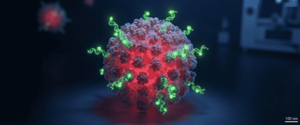
When evaluating nanoscale particle sizing against national benchmarks, laboratories frequently reference NIST particle metrology protocols to verify dimensional uniformity.
Colloidal Stability and Suspension Control
Sub-micron particles possess high surface energy, making them susceptible to aggregation if ionic strength or surfactant concentration fluctuates.
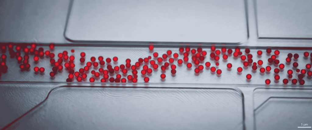
Utilizing optimized surfactant systems keeps individual spheres uniformly suspended, directly aligning with protocols in contamination control and manufacturing cleanroom standards governed by ISO standards body guidelines.
Overcoming Common Technical Challenges
Assay developers frequently encounter technical hurdles when transitioning from larger microparticles to 0.3µm sub-micron fluorescent standards. Addressing these obstacles requires precise control over buffer viscosity, wicking velocity, and blocking agents.
Mitigating Nonspecific Adsorption
Complex biological samples, such as human serum or plasma, often introduce matrix interference that elevates background fluorescence. Incorporating blocking agents like bovine serum albumin (BSA) or specialized detergents suppresses hydrophobic interactions on unassigned particle surface areas.
Documented methodologies in NCBI diagnostics research demonstrate that proper blocking significantly lowers baseline noise in low-abundance biomarker detection.
Technical Specification Matrix
| Parameter | Specification & Operational Value |
| Nominal Diameter | 0.3 µm (300 nm) monodisperse distribution |
| Surface Chemistry | Covalently bound, high-density streptavidin |
| Dye Architecture | Internally dyed core to eliminate fluorophore leaching |
| Excitation / Emission | Optimized for standard fluorophores (e.g., Europium / Green / Red) |
| Storage Requirements | 2°C to 8°C in original amber container (do not freeze) |
Reviewing advanced metrology guides assists engineers in calibrating optical readers to match the exact emission spectra of these microparticles. Furthermore, validating physical facility environments for sensitive assays relies on protocols detailed under cleanroom validation.
Advanced Applications in Modern Diagnostics
Biosensing platforms increasingly rely on sub-micron fluorescent reporters to enhance analytical sensitivity in point-of-care test kits and high-throughput automated analyzers.
Lateral Flow and Microfluidic Integration
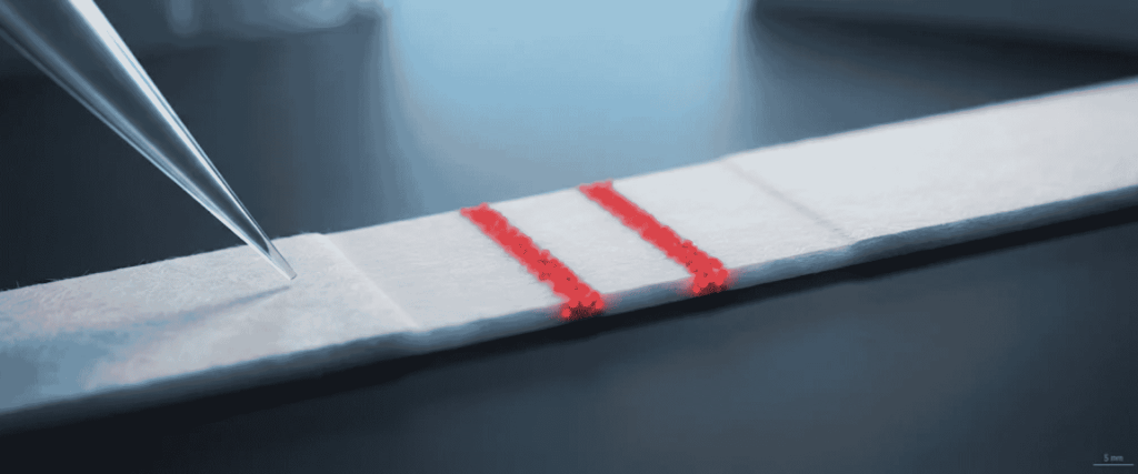
Integrating 0.3µm particles into nitrocellulose membranes optimizes capillary flow rates without causing membrane clogging or premature capture.
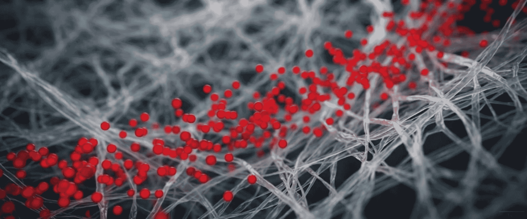
Peer-reviewed data published in PubMed biosensing studies confirm that smaller monodisperse reporter spheres dramatically improve capture kinetics compared to conventional micron-scale latex beads.
Conclusion
Optimizing 0.3µm Fluoro-Max streptavidin-coated particles requires rigorous control over internal dye stability, covalent surface binding, and buffer chemistry.
By addressing aggregation risks and eliminating fluorophore leaching, diagnostic developers can achieve exceptional sensitivity and reproducibility in clinical assays.
Partner with technical specialists to configure exact particle requirements for your specific validation and manufacturing workflow.
Frequently Asked Questions (FAQs)
1. What prevents dye leaching in Fluoro-Max particles?
Internal dye encapsulation seals fluorophores deep within the polymer matrix, isolating them from surrounding aqueous test buffers and preventing background signal degradation.
2. Why is 0.3µm selected for high-sensitivity LFIA strips?
A 0.3µm diameter balances high surface-area-to-volume ratio with optimal flow velocity through porous nitrocellulose membranes, preventing clogging while maximizing target capture sites.
3. How should streptavidin-coated particles be stored to preserve binding capacity?
Store particles between 2°C and 8°C in tightly sealed, upright containers protected from direct light. Freezing is strictly prohibited, as ice crystal formation disrupts colloidal stability and active binding sites.