
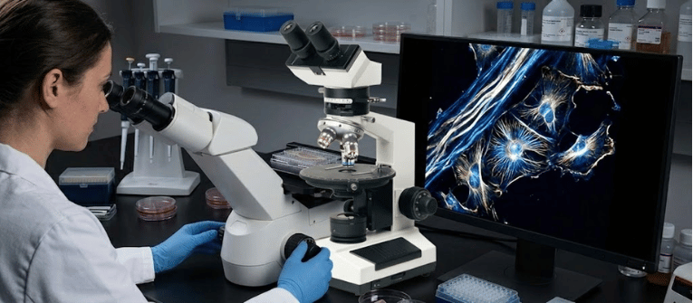
Conventional brightfield microscopes struggle to capture highly detailed images of transparent living cells without applying chemical stains.
While staining provides contrast, it often alters or destroys the living state of the biological sample.
Polarizing biological microscopy solves this issue by exploiting a physical property inherent to many native molecular structures: optical anisotropy or birefringence (Koike‐Tani et al., 2013).
By manipulating the vibration direction of light waves, these specialized optical systems reveal the hidden structural order within cellular components, tissues, and crystals while keeping the sample completely unaltered.
Most standard microscope illumination sources generate unpolarized light waves that vibrate in all directions perpendicular to the optical axis (Davidson, 2007).
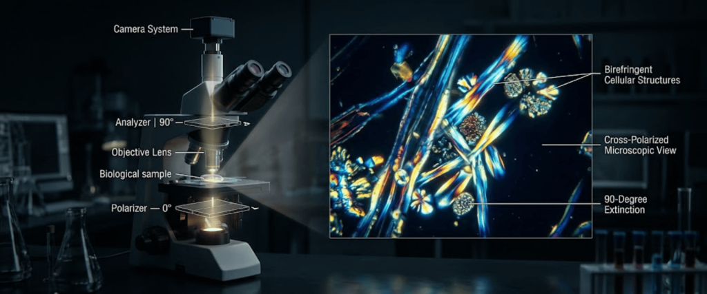
A polarizing biological microscope alters this path using two critical filters aligned orthogonally at a $90^\circ$ angle relative to one another:
When no sample is present, or when the light encounters an isotropic (monorefractive) substance, the polarized light passes through the specimen unchanged and is completely blocked by the analyzer.
This state is known as maximum extinction, resulting in a dark background image (Davidson, 2007; Koike‐Tani et al., 2013).
However, when the polarized light interacts with an anisotropic (birefringent) biological structure, the light wave splits into two distinct rays traveling at different velocities (Katoh et al., 1999; Koike‐Tani et al., 2013).
These rays recombine upon exiting the sample, changing the polarization state and enabling a portion of the light to successfully pass through the analyzer.
As a result, highly ordered biological structures stand out brightly against a dark backdrop without requiring artificial coloration (Koike‐Tani et al., 2013).
Polarizing microscopy is highly effective for identifying and quantifying structures featuring parallel, tightly aligned molecular bonds (Katoh et al., 1999).
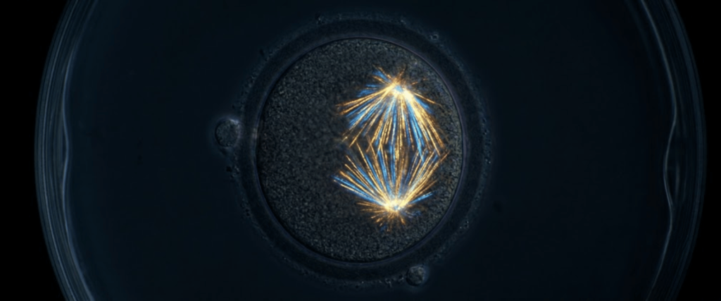
In assisted reproductive technology (such as IVF), assessing the density and alignment of the meiotic spindle in oocytes is a reliable, non-invasive method for evaluating cell viability before fertilization.
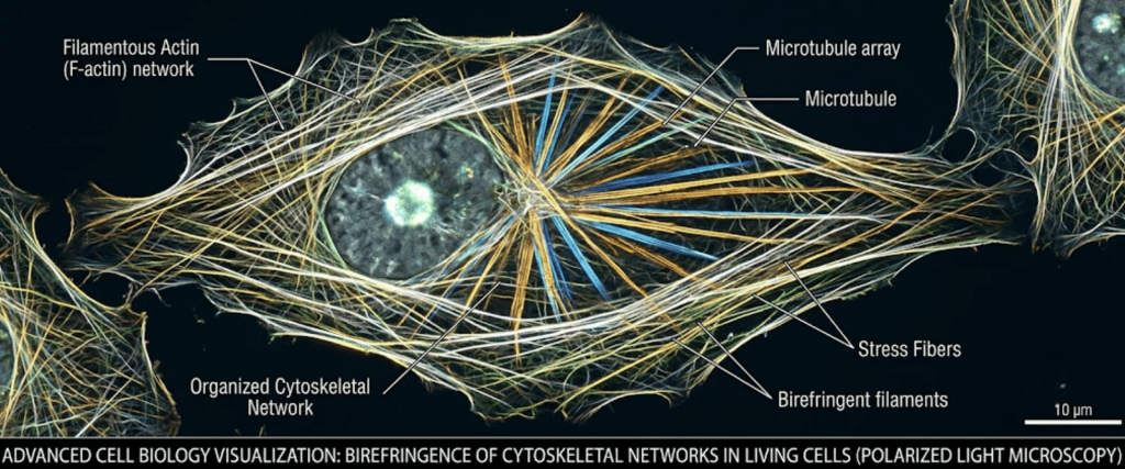
Highly organized polymers like filamentous actin and parallel microtubules show natural form birefringence, making them visible during real-time structural analysis (Katoh et al., 1999; Koike‐Tani et al., 2013).
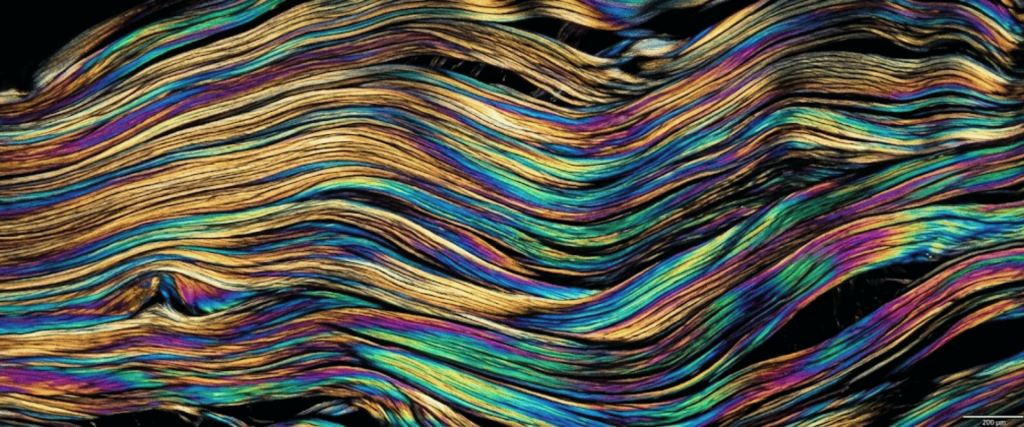
Structural proteins such as collagen form dense, organized arrays that exhibit distinct anisotropic properties when examined under polarized light (Koike‐Tani et al., 2013).
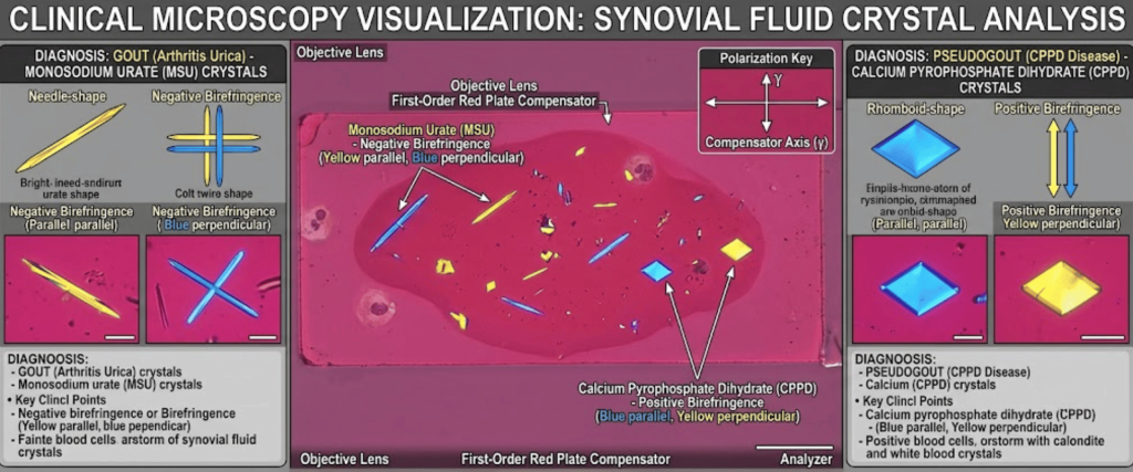
Clinical laboratories use this contrast mechanism to distinguish between monosodium urate (gout) crystals and calcium pyrophosphate (pseudogout) crystals in joint fluids based on their optical sign of elongation.
The table below highlights the practical differences between standard brightfield setups and specialized polarizing biological systems like the BMP-107 series.
| Feature / Metric | Standard Brightfield Microscope | Polarizing Biological Microscope (e.g., BMP-107B/T) |
|---|---|---|
| Primary Contrast Source | Light absorption variation through density or color | Phase shifts from optical anisotropy / birefringence |
| Optical Filters Required | None, except standard color or blue daylight filters | Fixed or sliding polarizer and rotatable analyzer |
| Objective Lens Design | Standard achromatic or plan achromatic lenses | Strain-free achromatic objectives: 4X, 10X, 40X |
| Stage Functionality | Fixed rectangular mechanical stage | revolving round stage with precise vernier scale |
| Specialized Optics | Not applicable | Bertrand lens, slip, and quartz wedges |
| Sample Preparation | Requires chemical dyes or stains for transparent samples | Label-free imaging of native, living structures |
To achieve accurate results, a polarizing biological microscope requires precise hardware modules designed to prevent external optical distortions.
Strain-Free Objectives: Standard glass lenses often contain internal stresses introduced during manufacturing. These stresses cause accidental birefringence that can distort image quality. Polarizing setups use specialized strain-free objectives to ensure background readings remain completely dark at maximum extinction (Koike‐Tani et al., 2013).
Polarizing biological microscopes offer high-contrast, label-free imaging for laboratories handling sensitive, highly structured samples.
By pairing crossed polarizers with strain-free optics, systems like the BMP-107 enable clear visualization of fine structural details from reproductive cells to industrial crystalline compounds without the downside of chemical staining.
The Bertrand lens changes the microscope’s view from standard imaging to an interference pattern view. This lets researchers examine directional light paths to calculate optical properties in crystals.
Standard lenses have internal manufacturing stresses that alter polarized light. Strain-free objectives prevent these optical distortions, keeping the background dark so users can see true sample birefringence.
Naturally birefringent structures include the meiotic spindle in eggs, collagen fibers in tissue, actin filaments in muscle, cell walls in plants, and various joint crystals.
Compensator plates introduce a known optical delay to the light path. This turns subtle phase changes into distinct colors, making it easier to see and measure faint biological details.